Their health
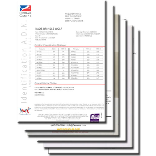
We are very demanding on the health and we update « bad » and « good » health results.
Most of the diseases are multifactorial (genetic, hereditary, food, environment, unknown, etc.) and not the fault of the owner nor the breeder!
As we know, “prevention is better than cure”!
That’s why we highly test our dogs for every disease possible at our disposal:
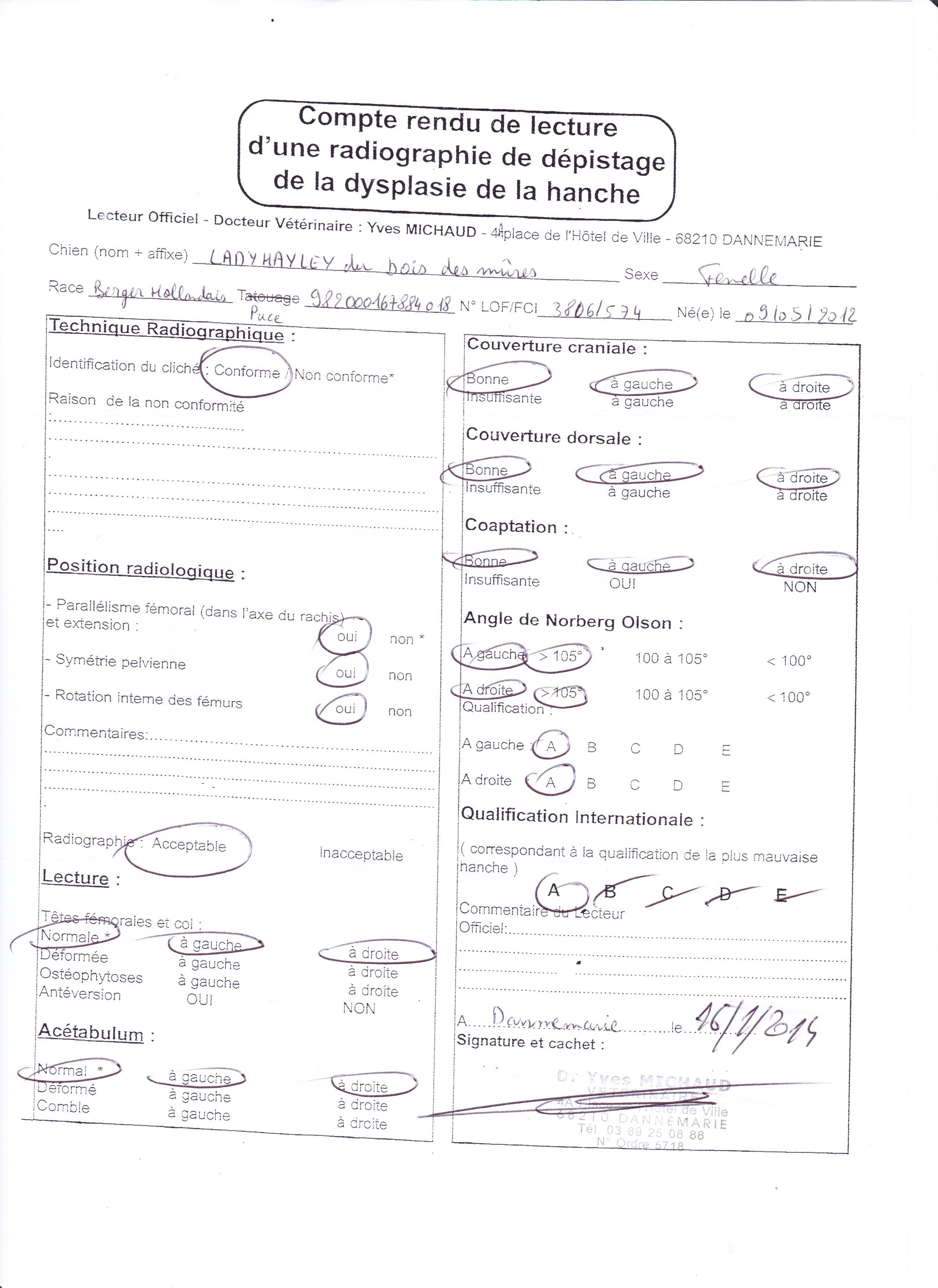
French official xrays for:
- HD (hips dysplasia),
- ED (elbows dysplasia),
- SP (spondylosis dissecans),
- OCD (osteochondrosis dissecans).
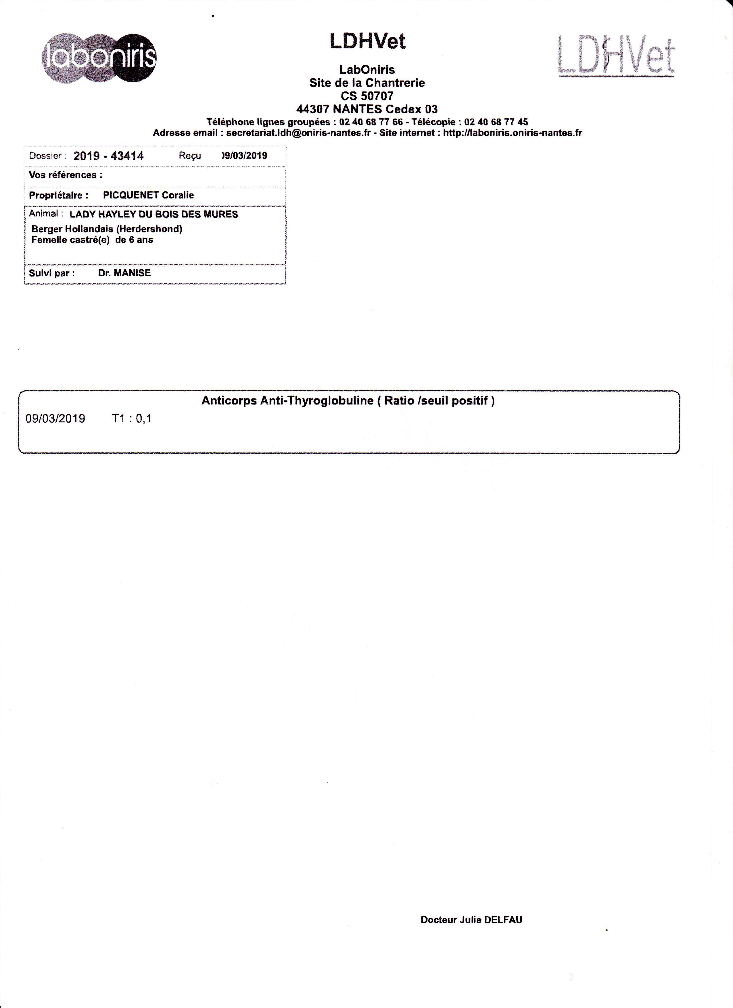
Blood samples for:
- TgAA (hypothyroidism) every year.
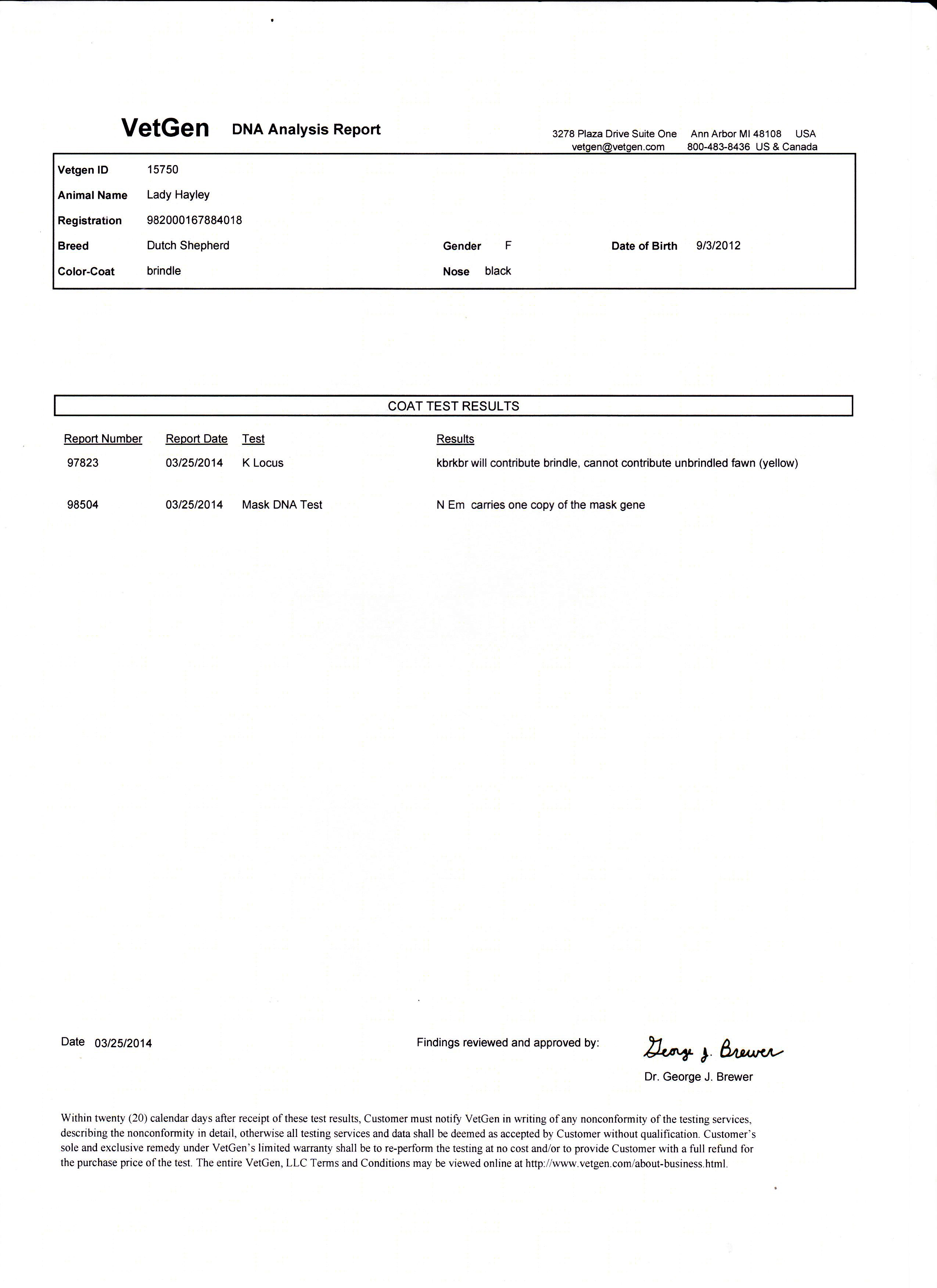
DNA tests for:
- DNA with parentage from the Société Centrale Canine,
- Kbr-locus (brindle) from VetGen lab,
- D-locus (dilution), from GenSol lab,
And with MyDogDNA panel tests including:
- vWD1 (von Willebrand disease type I),
- DM (degenerative myelopathy),
- MDR1 (multi-drug resistance 1),
- SDCA 1 & 2 (spongy cerebellar degeneration with cerebellar ataxia),
- Em-locus (mask).
We go to the osteopath regularly.
Hips dysplasia
source: AKC.ORG
Hip dysplasia is a common skeletal condition, often seen in large or giant breed dogs, although it can occur in smaller breeds, as well.
To understand how the condition works, owners first must understand the basic anatomy of the hip joint.
The hip joint functions as a ball and socket. In dogs with hip dysplasia, the ball and socket do not fit or develop properly, and they rub and grind instead of sliding smoothly.
This results in deterioration over time and an eventual loss of function of the joint itself.
Several factors lead to the development of hip dysplasia in dogs, beginning with genetics. Hip dysplasia is hereditary and is especially common in larger dogs.
Factors such as excessive growth rate, types of exercise, and improper weight and nutrition can magnify this genetic predisposition.
Some puppies have special nutrition requirements and need food specially formulated for large breed puppies. These foods help prevent excessive growth,
which can lead to skeletal disorders such as hip dysplasia, along with elbow dysplasia and other joint conditions. Slowing down these breeds’ growth
allows their joints to develop without putting too much strain on them, helping to prevent problems down the line.
Improper nutrition can also influence a dog’s likelihood of developing hip dysplasia, as can too much exercise – or too little.
Obesity puts a lot of stress on your dog’s joints, which can exacerbate a pre-existing condition such as hip dysplasia or even cause hip dysplasia.
Talk to your vet about the best diet for your dog and the appropriate amount of exercise your dog needs each day to keep them in good physical condition.
Elbows dysplasia

source: ACVS.ORG
Elbow dysplasia is a condition involving multiple developmental abnormalities of the elbow joint.
The elbow joint is a complex joint made up of 3 bones (radius, ulna, and humerus). If the 3 bones do not fit together perfectly due to growth abnormalities,
abnormal weight distribution on areas of the joint occur causing pain, lameness, and the development of arthritis.
The cause of ED in dogs remains unclear. There are a number of theories as to the exact cause of the disease that include genetics, defects in cartilage growth, trauma, diet, and so on.
It is most commonly suspected this is a multifactorial disease in which causes the growth disturbances.
Unfortunately, once the elbow joint has been damaged through either cartilage loss, medial compartment disease or an ununited anconeal process, inflammation and further cartilage damage
occurs. Ultimately this causes progressive arthritis of the elbow joint leading to pain and loss of function.
Spondylosis
source: VCAHOSPITALS.COM
Spondylosis deformans is a condition that affects the vertebral bones of the spine and is characterized by the presence of bony spurs or osteophytes along the edges of the bones
of the spine. A bony spur may develop in a single spot on the spine; more commonly, there will be multiple bone spurs in several different locations along the spine.
The most common places that spondylosis deformans lesions develop are along the thoracic vertebrae (chest), especially at the junction between the rib cage and the abdomen, in the
lumbar spine (lower back) and in the lumbosacral spine (around the hips and back legs). In some cases the bony spurs may become large enough that they appear to form a complete
bridge between adjacent vertebral bones.
Spondylosis deformans is a chronic condition that is associated with aging. Research indicates that it often develops as a secondary problem related to degenerative disease of the
intervertebral discs.
Osteochondrosis
source: ACVS.ORG
Osteochondrosis occurs commonly in the shoulders of immature, large, and giant-breed dogs. The lesion usually appears on the caudal (back) surface of the humeral head.
Osteochondrosis begins with a failure of immature cartilage to form bone in the humeral head. This failure leads to abnormal cartilage thickening. Increased cartilage thickness
may result in malnourished cartilage cells that die. Loss of these cartilage cells deep in the cartilage layers leads to formation of a defect at the junction between cartilage and bone.
Subsequently, normal daily activity may cause fissures in the cartilage that eventually communicate with the joint, forming a cartilage flap. It is with the formation of a flap
that osteochondrosis becomes osteochondritis dissecans (OCD).
OCD is the form of osteochondrosis that is associated with pain and dysfunction. In some cases, the resulting flap occupies as
much as half the humeral head. The cartilage flap may completely detach from the underlying bone and become lodged in the back of the joint pouch. Free cartilage flaps can lodge in joints
and may increase in size with calcification becoming "joint mice” which can be seen on radiographs.
Hypothyroidism
source: VETMED.WSU.EDU
The most common signs of low thyroid function in dogs include loss or thinning of the fur, dull hair coat, excess shedding or scaling, weight gain, reduced activity and reduced ability to tolerate the cold. The hair loss occurs primarily over the body, sparing the head and legs, and is usually not accompanied by itching or redness of the skin. Some dogs will have thickening of the skin and increased skin pigment, especially in areas of friction, such as the armpit (axilla). Hypothyroid dogs often have ear infections and show ear pain, redness, and odor. Hypothyroid dogs may also develop skin infections which may be itchy and result in sores on the body. The accumulation of substances called mucopolysaccharides can cause the muscles of the face to droop giving the dog a facial expression that is sometimes called “tragic”.
Less commonly recognized signs that may be seen in a small number of dogs with hypothyroidism include dilation of the esophagus (megaesophagus) causing regurgitation, and abnormal function of nerves or muscles leading to weakness or abnormal ability to walk.
Routine blood tests can be affected by hypothyroidism, although the changes are not consistent and can be subtle. Dogs may have a mild anemia and increased levels of cholesterol.
Kbr locus
source: VETGEN.COM
Kbr is dominant only to ky. kbr is responsible for the brindle trait and for a long time had been considered to belong in the E locus. Recent breeding studies had also shown this to be incorrect. The recessive allele, ky, allows the basic patterns of the A locus to be expressed. So too does the kbr allele, but with brindling of any tan, fawn, or tawny areas.
kbr kbr allows A locus to express (tan point, tricolor, fawn, sable, tawny) with brindling
kbr ky allows A locus to express (tan point, tricolor, fawn, sable, tawny) with brindling
ky ky allows expression of agouti patterns without brindling
D locus
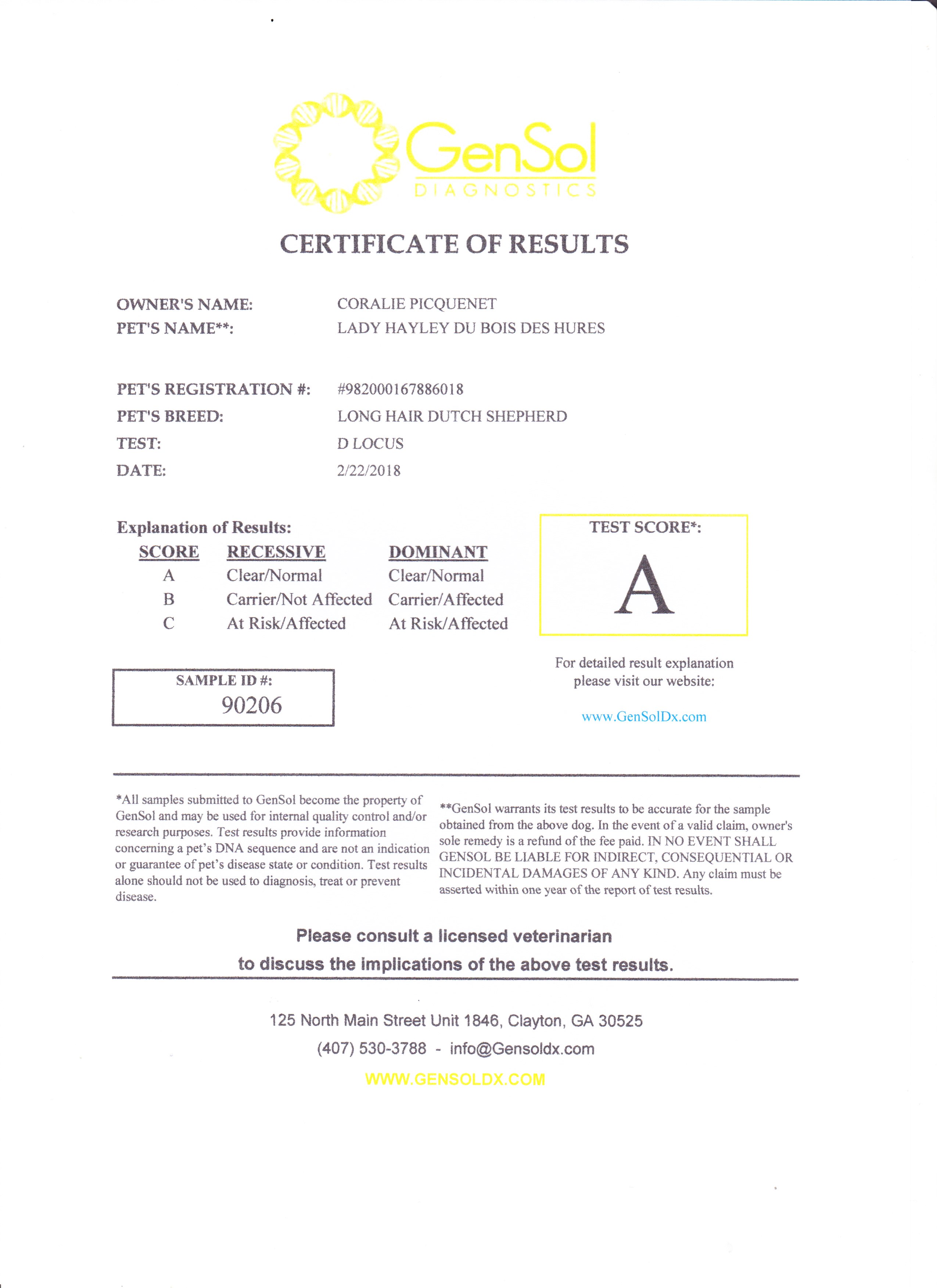
source: VETGEN.COM
The D locus is the primary locus associated with diluted pigment, which results in coats that would otherwise be black or brown instead showing up as gray, or blue in the case of black,
and pale brown or Isabella in the case of brown.
The melanophilin gene has recently been shown to be responsible, but not all of the dilute causing mutations have been identified yet.
Von Willebrand type I
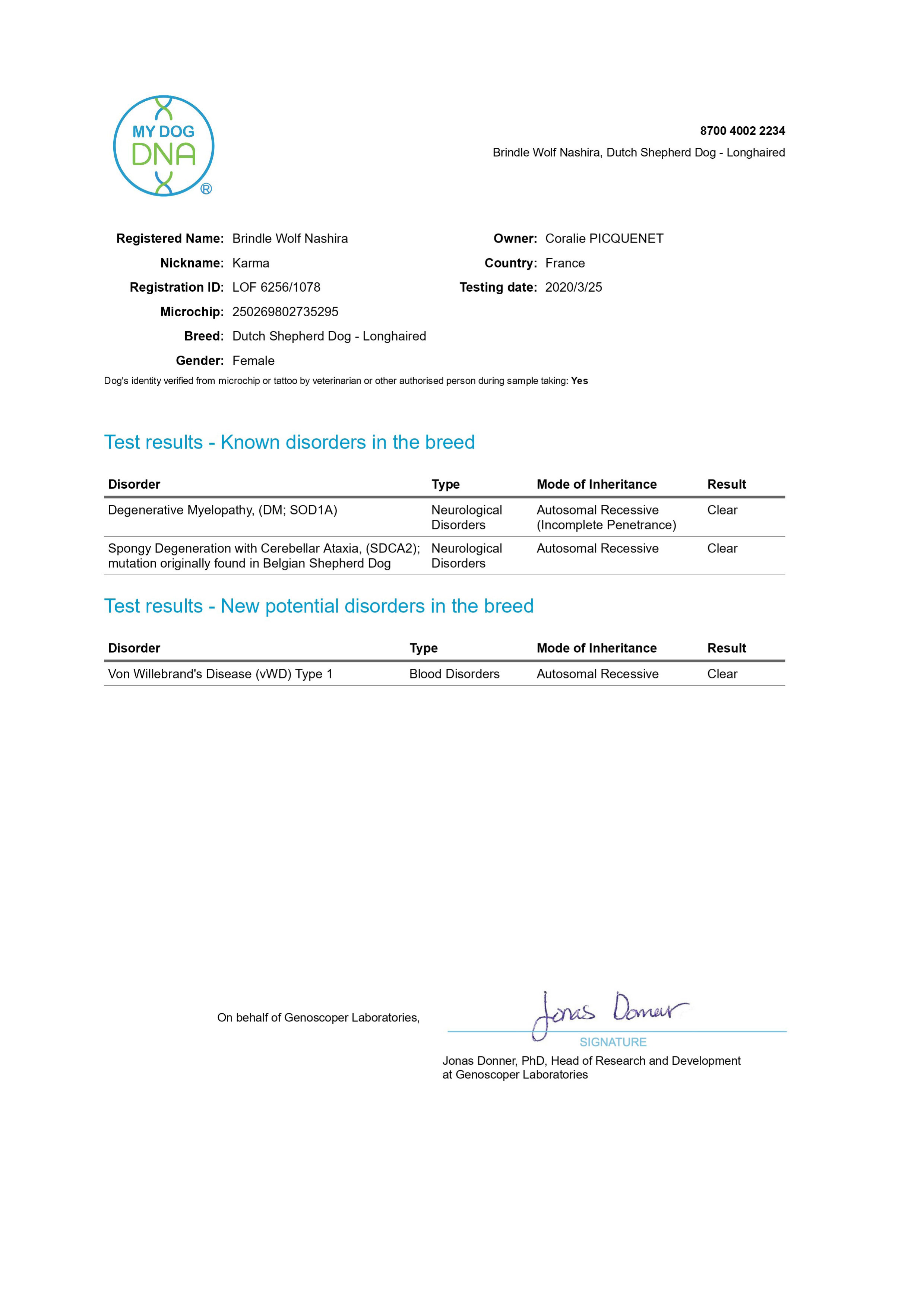
source: MYDOGDNA.COM
The clotting pathway is a complex process. The vWF glycoprotein complexes with factor VIII and both are required for platelet adhesion and preventing the rapid clearance of factor VIII.
Von Willebrand’s disease (vWD) is a bleeding disorder affecting multiple breeds and several genetic variants have been characterised. Type 1 is the mildest form of vWD in which the level
of von Willebrand’s factor is reduced though all the multimers are present. Many cases are subclinical but can be associated with an increased bleeding tendency after surgery or trauma.
The disease is inherited in an autosomal recessive manner, though some carriers can have clinical signs.
Degenerative Myelopathy

source: MYDOGDNA.COM
Degenerative myelopathy (DM) is an inherited neurological disorder found in many dog breeds.
It is not yet clear if all dogs carrying two copies of the mutation will develop clinical signs,
especially considering the variable presentation noted amongst breeds found to carry it. Clinical signs develop at an old age and all affected dogs do not develop symptoms during their
natural lifespan. It is unclear, whether the disease process is ongoing in these dogs or if the mutation does not cause disease in all affected dogs.
DM is inherited in an autosomal recessive fashion.
Multi-Drug Resistance 1
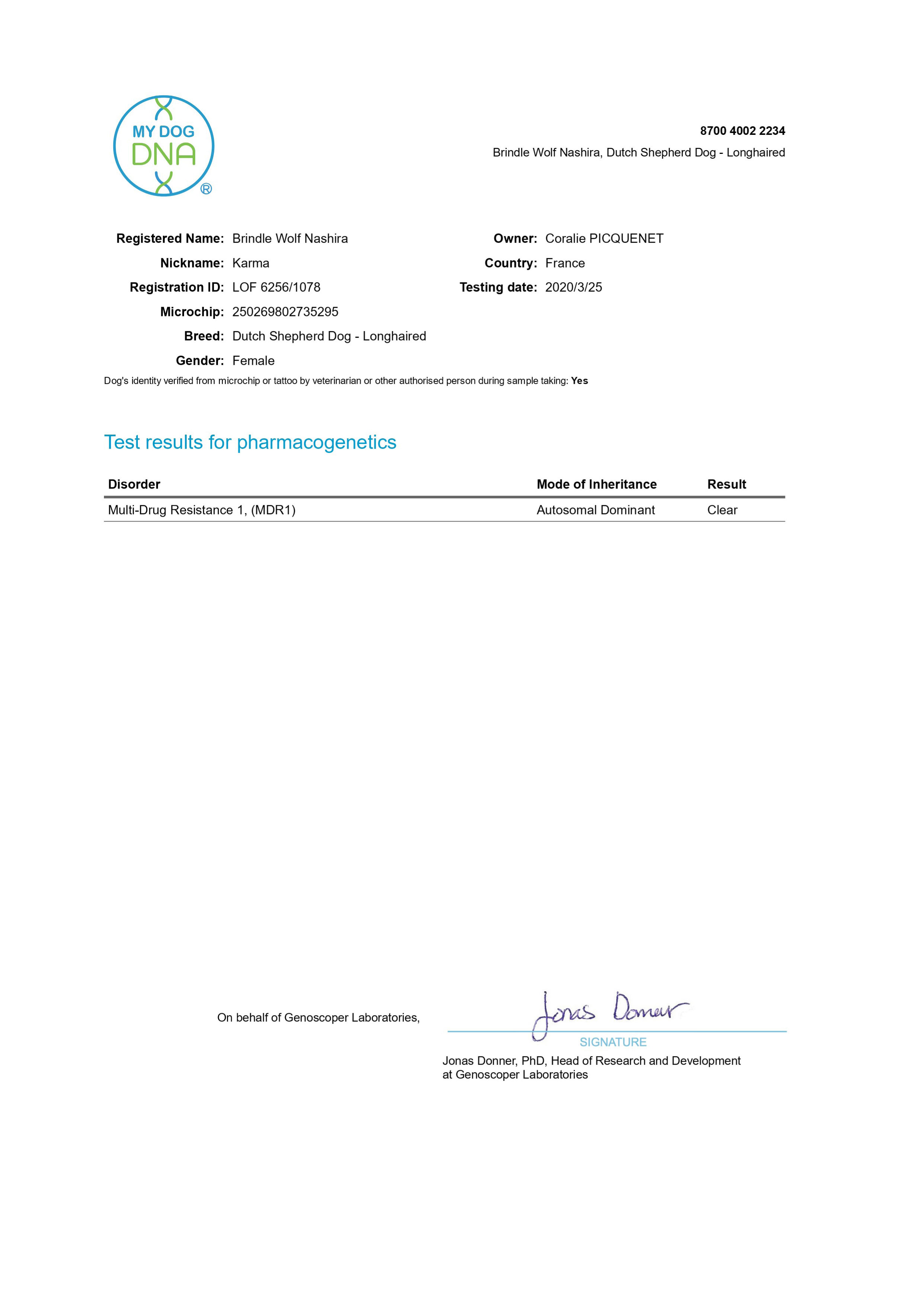
source: MYDOGDNA.COM
Multi-drug resistance 1 is a genetic mutation that alters a dog's ability to limit the absorption and distribution of many drugs.
Affected dogs are slower to eliminate drugs from the body and can suffer side effects when exposed to certain medications. This mutation is sometimes also called "ivermectin sensitivity".
However, the name is a misnomer as several other drugs pose a risk to MDR1 positive dogs. Adverse reactions can occur when affected dogs are exposed to some common drugs such as acepromazine, butorphanol, and macrocyclic lactones.
However, all FDA approved heartworm preventatives are safe to administer to MDR1 positive dogs.
This mutation is inherited in a dominant fashion though dogs with two copies of the mutation will exhibit more severe clinical signs.
Spongy degeneration with cerebellar ataxia

source: MYDOGDNA.COM
SeSAME/EAST is a rare hereditary neurological disorder encountered in humans. Belgian Shepherds, especially the Malinois type, can suffer from a syndrome that is similar to this human
condition. This syndrome in Belgian Shepherds is called Spongy Degeneration with Cerebellar Ataxia. The inheritance pattern is autosomal recessive.
The age of onset of spongy degeneration with cerebellar ataxia is usually 4-8 weeks. The first observable sign is poor coordination of movements (ataxia) and generalized cerebellar
dysfunction. Affected dogs may also suffer from episodic seizures and demonstrate pacing and circling behaviour.
Disease progression is rapid and often results in euthanasia over welfare concerns.
Em locus
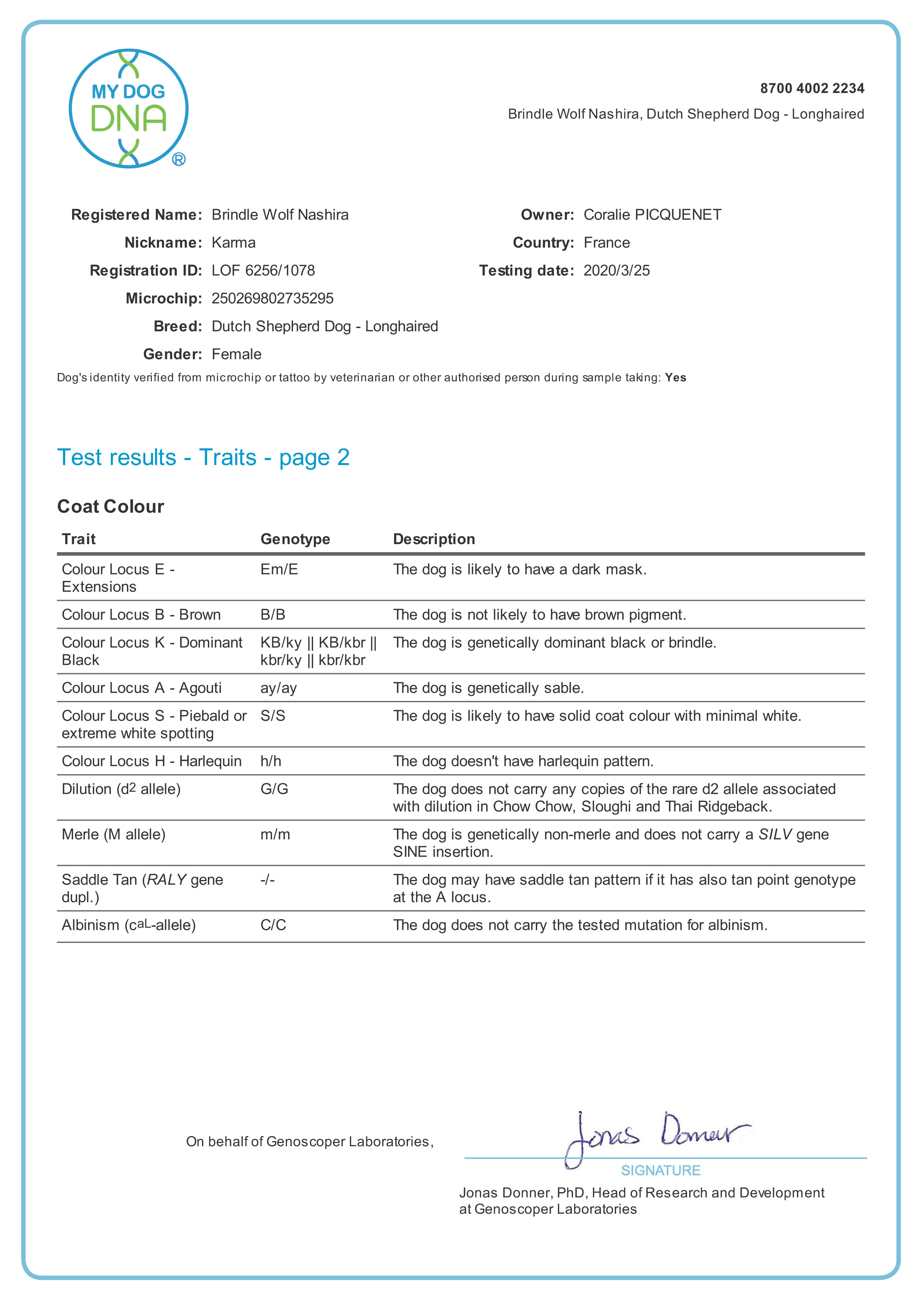
source: VETGEN.COM
This analysis reveals whether a dog with a mask has one or two copies of this version of the extension locus. Animals with a single copy can produce offspring with or without a mask,
while those with two copies will only produce masked offspring. The test may also be applied to black dogs where it may not be possible to tell if there is a mask.